The calcaneofibular ligament has distinct anatomic
morphological variants: an anatomical cadaveric study
Bruno S. Pereira, C. Niek van Dijk, Renato Andrade, Ricardo P. Casaroli-Marano, João Espregueira-Mendes, Xavier Martin Oliva
What did the study consist of?
In this study we performed anatomical dissection to evaluate the morphology and morphometry of the calcaneofibular ligament.
And what results have been achieved?
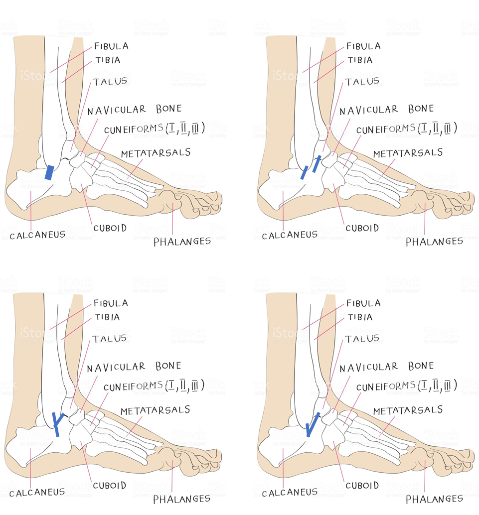
- After dissecting 47 cadaveric ankle specimens we found that the calcaneofibular ligament demonstrates 4 variations:
- single beam (44.7%)
- double beam (10.6%)
- Y-format double beam (27.7%)
- V-shaped double beam (17.0%)
- beam length was higher in the single-beam and double-beam Y-shaped variants.
- The size of the anatomical footprints of origin and insertion of the ligament were heterogeneous when comparing the different anatomical variations.
What is the clinical relevance?
- It is still necessary to understand what the relationship of these variants of the calcaneofibular ligament are to the lateral astragalo-calcaneal ligament and the inferior fascicle of the anterior peroneo-astragalineus.
- Information about the variants found (morphologies) and the measurements taken (morphometry) can be used to improve the diagnostic process and surgical approach (choice and size of the graft to be used, as well as in placing the graft in the anatomical position).
- This remains to be studied in future work:
- Does the double beam provide greater subtalar stability?
- Could double bundle reconstruction of the peroneal-calcaneal ligament postoperatively result in a lower recurrence rate of sprains with increased ankle stability and functionality?
This article can be found in the KSSTA journal at the link below:
https://link.springer.com/article/10.1007%2Fs00167-019-05797-5
